BLOOD POISONING Staphylococcus bacteria
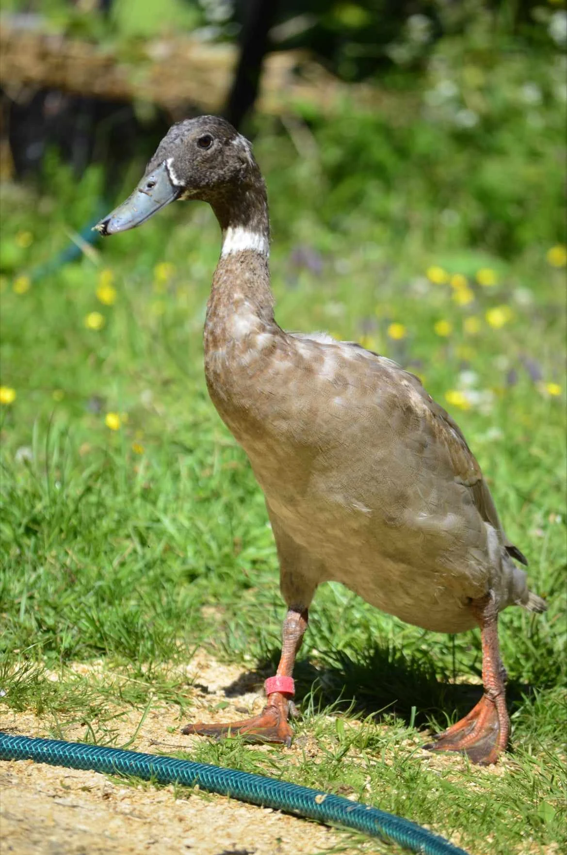
Dumptruck
3 years old, Indian Runner duck
Everything happened so quickly. When he first began showing signs of illness, it only took a few days before he passed away. It started with him separating himself from the flock, followed by him spending more and more time lying down. He stopped eating, and when I weighed him, I discovered that he had lost 400 grams in less than a month. On the day he died, he lost the ability to walk. Later that evening, when I picked him up, he finally seemed to relax—and he passed away peacefully in my arms.
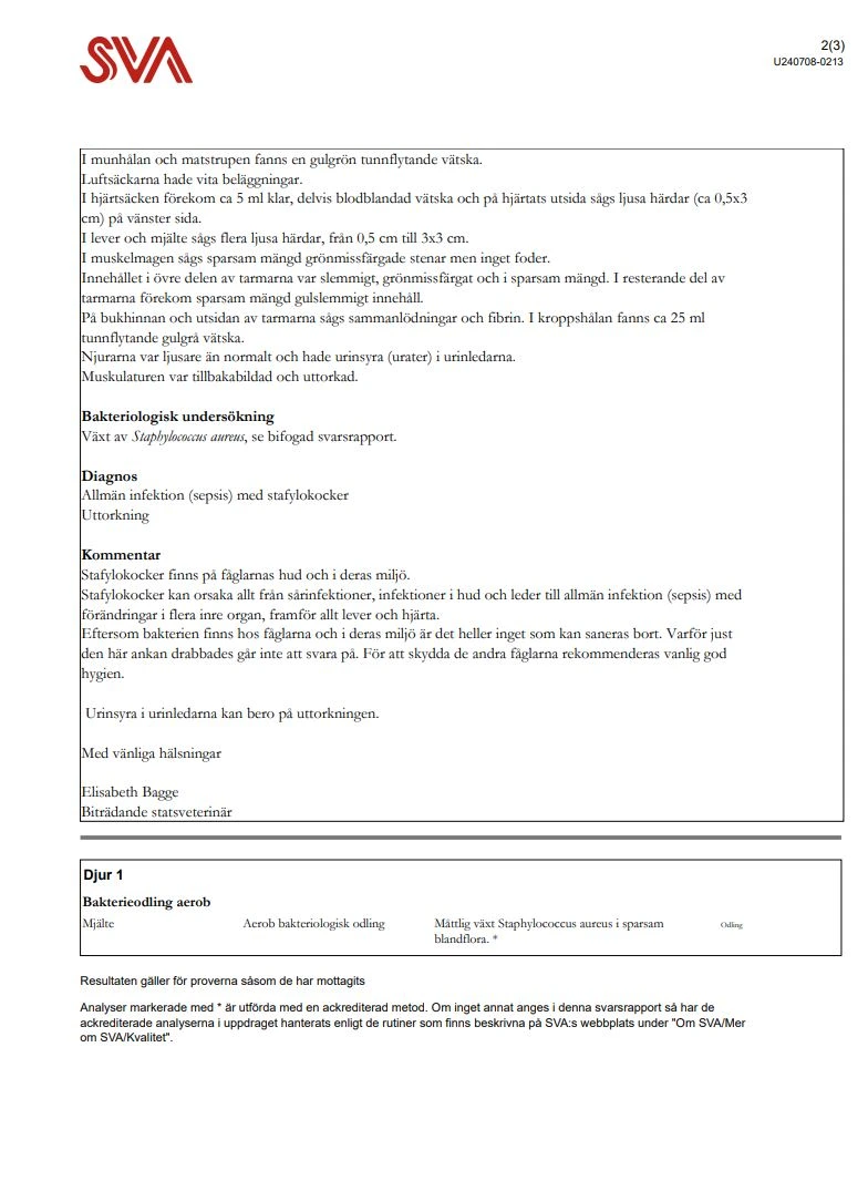
I sent him for a post-mortem examination, which revealed the extent of the damage throughout his body. He had lesions in multiple organs and tissues. A bacterial culture confirmed Staphylococcus aureus as the cause, and it is highly likely that he had developed sepsis, as the disease progressed so rapidly.
Dumptruck had already suffered from an eye infection during his first year of life, caused by bacteria that were likely already present within his own body. Even at that time, this suggested that he may have had a weakened immune system, making him particularly vulnerable to a severe staphylococcal infection.
2020/12/24 – 2024/07 /06

English Translation
In the oral cavity and esophagus, a yellowish-green, thin fluid was found. The air sacs had white coatings. In the pericardium, there was approximately 5 ml of clear, partially blood-tinged fluid, and on the outside of the heart, pale lesions (approx. 0.5×3 cm) were seen on the left side. In the liver and spleen, several pale lesions were seen, ranging from 0.5 cm to 3×3 cm. In the gizzard, a sparse amount of green-discolored stones was seen, but no feed. The content in the upper part of the intestines was slimy, greenish-gray, and present in sparse amounts. In the remaining part of the intestines, a sparse amount of yellowish-slimy content was present. On the peritoneum and the outside of the intestines, adhesions and fibrin were seen. In the body cavity, there was approximately 25 ml of thin, yellowish fluid. The kidneys were paler than normal and had uric acid (urates) in the ureters. The musculature was atrophied (wasted) and dehydrated.
Bacteriological examination
Growth of Staphylococcus aureus, see attached response report.
Diagnosis
Systemic infection (sepsis) with staphylococci Dehydration
Comment
Staphylococci are found on the birds’ skin and in their environment. Staphylococci can cause everything from wound infections, skin and joint infections to systemic infection (sepsis) with changes in several internal organs, especially the liver and heart. Since the bacteria are present on the birds and in their environment, it is also nothing that can be eradicated. Why this particular duck was affected is impossible to answer. To protect the other birds, standard good hygiene is recommended.
Uric acid in the ureters may be due to the dehydration.
Best regards,
Elisabeth Bagge Assistant State Veterinarian
Animal 1
Aerobic bacterial culture
-
Spleen: Aerobic bacteriological culture — Moderate growth of Staphylococcus aureus in a sparse mixed flora. * (Culture)
